
Abstract
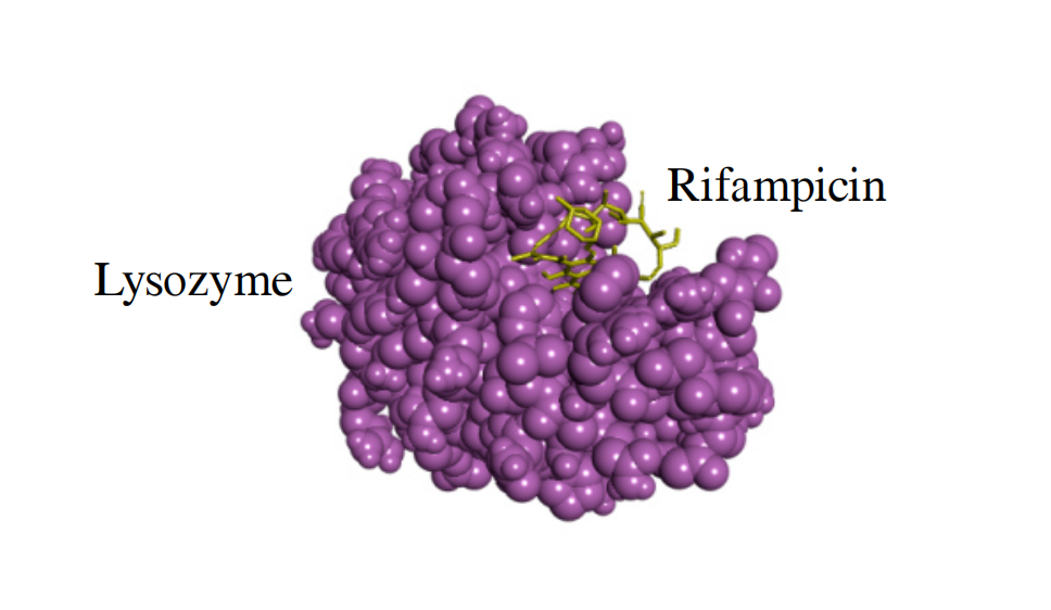
Hen egg-white lysozyme interacts with rifampicin through van der Waals interactions, forming complexes in which one or two molecules of the antibiotic bind to a single lysozyme molecule. The complex formation does not involve the active site of lysozyme, thereby enabling further investigation of these complexes in cellular and animal models.
Keywords
Communication
Molecular medicine has become an integral component of the modern healthcare. In biological fluids, most drugs exist either as free molecules or in complexes with biomolecules such as proteins. Protein binding influences drug distribution in tissues and organs, metabolism, and clearance from the body. Furthermore, protein–drug interactions can modulate drug delivery rates, toxicity, and pharmacological efficacy.[1-5]
Lysozyme is of considerable biological importance not only as an antimicrobial enzyme but also as a well-characterized model protein.[6] Investigating interactions between lysozyme and antibiotics may improve our understanding of antibiotic action and facilitate the development of enzyme–antibiotic complexes for therapeutic applications.[7] Rifampicin is a key component of anti-tuberculosis therapy. The antibiotic specifically inhibits the DNA-dependent RNA polymerase in Mycobacterium tuberculosis.[8] Lysozyme hydrolyzes the β-1,4-glycosidic bonds between N-acetylglucosamine (GlcNAc) and N-acetylmuramic acid (MurNAc) in the peptidoglycan of cell walls of pathogenic microorganisms, inducing cell lysis. Glycosidic bonds between GlcNAc and MurNAc are components of the mycobacterial cell wall.[9, 10] Combining antimicrobial agents with different mechanisms of action seems to be the most important step in the fight against M. tuberculosis and other pathogens. Here, the interaction between hen egg-white lysozyme and rifampicin was investigated using fluorescence spectroscopy and molecular docking.
Lysozyme–rifampicin complexes were prepared in phosphate-buffered saline (PBS), with rifampicin initially dissolved in dimethyl sulfoxide (DMSO). The final solutions contained 17 μmol dm−3 of lysozyme and 0.5-, 1-, 2-, and 4-fold molar excesses of rifampicin, while the DMSO concentration did not exceed 2.5 vol%. The detailed description of the reagents and methods is provided in Online Supplementary Materials.
Figure 1(a) shows the fluorescence spectra of lysozyme alone and in the presence of rifampicin at 25 °C. The fluorescence spectrum of lysozyme displayed a characteristic maximum at 345 ± 2 nm. Increasing rifampicin concentration resulted in progressive quenching of lysozyme fluorescence and a shift of the fluorescence maximum toward longer wavelengths (red shift or bathochromic shift). At the temperatures of 33, 40, and 50 °C, the changes in the lysozyme fluorescence spectrum under the influence of rifampicin were similar to those at 25 °C (Figure S1, Online Supplementary Materials). It may be concluded that antibiotic molecules influence the lysozyme molecules and that there is an interaction between them.
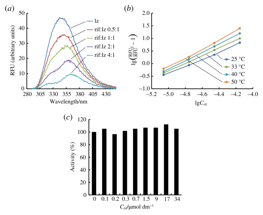
In the lysozyme structure, two tryptophan residues, Trp62 and Trp108, are located on the protein surface and contribute approximately 80% of the fluorescence signal.[11] A shift in the position of the lysozyme fluorescence maximum to longer wavelengths indicates an increase in the polarity of the fluorophore microenvironment (Trp62 and Trp108) when influenced by rifampicin. The increase in the polarity of Trp62 and Trp108 residues is likely due to a conformational change in the lysozyme molecule. This conformational change may bring the Trp residues closer to charged groups.
BSA, tyrosinase, and α-glucosidase have shown changes in their protein fluorescence spectra (quenching and a shift of the maximum toward longer wavelengths) depending on the amount of rifampicin added.[12-14] According to the researchers, the obtained data can be explained by conformational changes in protein molecules, resulting from the formation of complexes between them and rifampicin. Therefore, our data suggest that hen egg-white lysozyme and the antibiotic rifampicin form a complex. In order to assess the binding parameters of lysozyme to rifampicin, the temperature dependence of lysozyme fluorescence quenching was analyzed.
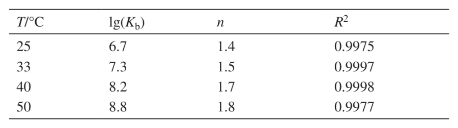
The dependences of lysozyme fluorescence intensity (λem = 345 nm) of rifampicin concentration are linearized (Figure 1(b)) in coordinates of the double-logarithmic Stern–Volmer equation (see Online Supplementary Materials), from which the values of Kb (the binding constant of lysozyme with rifampicin) and n (the number of binding sites) can be determined. The effective values of the parameters of lysozyme binding to rifampicin are presented in Table 1. It is evident that as the temperature increases, the number of binding sites n increases from 1.4 to 1.8. It can be assumed that rifampicin molecules are statistically distributed among lysozyme molecules. There are one or two antibiotic molecules per one molecule of enzyme. With increasing temperature, the number of lysozyme molecules bound to two antibiotic molecules increases, and the number of lysozyme molecules bound to one rifampicin molecule decreases. It is possible that with increasing temperature the number of collisions between enzyme and antibiotic molecules increases, which leads to an increase in the n parameter. The Kb values vary in order of magnitude at different temperatures, and an increase in lg(Kb) is observed with increasing temperature (Table 1). The Gibbs free energy of lysozyme binding to rifampicin (ΔG°) is −35 kJ mol−1, indicating the spontaneous interaction between the protein and the antibiotic.
The biological function of lysozyme is the hydrolysis of chemical bonds in the peptidoglycan of bacterial cell walls. Exploration of the effect of rifampicin on lysozyme activity is relevant from both scientific and practical perspectives. Rifampicin has been shown to inhibit the activity of certain enzymes, such as tyrosinase and α-glucosidase.[12, 13] Figure 1(c) shows the dependence of lysozyme activity on the concentration of rifampicin in the reaction medium (with Micrococcus lysodeikticus substrate). Figure 1(c) shows that lysozyme activity remains unchanged at 10- to 200-fold molar excesses of rifampicin. Since lysozyme is a component of saliva, blood, and other biological fluids, maintaining its biological function at large excesses of rifampicin is important.
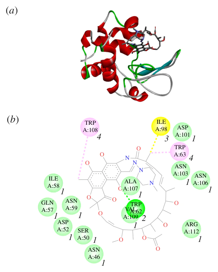
The structure of the lysozyme–rifampicin complex with the minimal ΔG° value is shown in Figure 2(a). For simulation, the lysozyme structure was taken from the Protein Data Bank (PDB ID 6LYZ). The rifampicin structure was generated and then optimized in the MM2 force field using the ChemOffice software package (ChemDraw, Chem3D). Blind docking was performed using Autodock 4.2, followed by visualization of the results in Discovery Studio (version 4.5). Figure 2(a) shows that the rifampicin molecule is located on the surface of the lysozyme molecule, where it is retained by non-covalent interactions. The main type of interaction determining the binding of lysozyme to rifampicin is nonspecific van der Waals interactions. For the lysozyme–rifampicin complex, van der Waals forces are identified between the antibiotic and amino acid residues Ala107, Val109, Ile58, Gln57, Asn59, Asp52, Ser50, Asn46, Asp101, Asn103, Asn106 and Arg112. The specific interaction forces involved in the binding of rifampicin with lysozyme are hydrophobic interactions and hydrogen bonding. Rifampicin produces hydrophobic interactions with Ile98, Trp63, and Trp108 and forms a hydrogen bond with Trp62 (Figure 2(b)). Our results demonstrate that rifampicin does not affect the activity of lysozyme at a 10- to 200-fold molar excess of the antibiotic (Figure 1(c)). It can be assumed that the antibiotic molecule is not complementary to the surface of the enzyme molecule. For this reason, a large number of specific interactions between lysozyme and rifampicin cannot be formed. It is known that the enzymatic activity of lysozyme is defined by two amino acid residues within its active site: Glu35 and Asp52.[15] Asp52 is involved in the interaction with rifampicin, but the very weak van der Waals force does not affect the ability of the enzyme to destruct the bacterial cell wall. The interaction between rifampicin and surface amino acid residues Trp108 and Trp62 of the lysozyme molecule can explain the quenching of the enzyme fluorescence by the antibiotic. The formation of the complex between the enzyme and the antibiotic is characterized by ΔG° = −20 kJ mol−1. The discrepancy between the calculated ΔG° value and the theoretical one may be due to a number of limitations of molecular docking (protein molecule rigidity; the solvent is excluded; failure to take into account pH and protein and ligand concentrations, etc.).
In summary, hen egg-white lysozyme and rifampicin spontaneously form stable complexes. Fluorescence spectroscopy demonstrated that one lysozyme molecule can bind one or two rifampicin molecules, depending on temperature. Molecular docking revealed that the interaction is mediated primarily by van der Waals forces. The complex formation does not involve the active site of lysozyme and therefore does not impair its enzymatic activity. These findings provide a foundation for further research into the antimicrobial properties of lysozyme–rifampicin complexes in cellular and animal models and highlight their potential biomedical applications. Modeling studies of the structure of lysozyme–rifampicin complexes at high temperatures are perspective and need the further support.
This research was funded by the state registration task (project 123032300028-0).
References