
Home / Articles / Human blood antibodies recognizing Galβ1-3GlcNAcα epitope in bacterial polysaccharide

Human blood antibodies recognizing Galβ1-3GlcNAcα epitope in bacterial polysaccharide

S. V. Tsygankova 1, 
S. V. Tsygankova
P. S. Obukhova 1, 2, 
P. S. Obukhova
A. V. Perepelov 3, 
A. V. Perepelov
N. V. Shilova 1, 2, 
N. V. Shilova
G. V. Pazynina 1, 
G. V. Pazynina
Published 7 October 2026 , received 25 June 2026
Communication
0
Cite this
GOST
 | 
Cite this
GOST
Tsygankova S. V. et al. Human blood antibodies recognizing Galβ1-3GlcNAcα epitope in bacterial polysaccharide // Mendeleev Communications. 2026.
GOST all authors (up to 50)
Tsygankova S. V., Obukhova P. S., Perepelov A. V., Shilova N. V., Paramonov A. S., Pazynina G. V., Bovin N. V. Human blood antibodies recognizing Galβ1-3GlcNAcα epitope in bacterial polysaccharide // Mendeleev Communications. 2026.
RIS
 | 
Cite this
RIS
TY - JOUR
DO - 10.71267/mencom.8127
UR - https://mendcomm.colab.ws/publications/10.71267/mencom.8127
TI - Human blood antibodies recognizing Galβ1-3GlcNAcα epitope in bacterial polysaccharide
T2 - Mendeleev Communications
AU - Tsygankova, S. V.
AU - Obukhova, P. S.
AU - Perepelov, A. V.
AU - Shilova, N. V.
AU - Paramonov, A. S.
AU - Pazynina, G. V.
AU - Bovin, N. V.
PY - 2026
DA - 2026/10/07
PB - Mendeleev Communications
ER -
BibTex
Cite this
BibTex (up to 50 authors)
@article{2026_Tsygankova,
author = {S. V. Tsygankova and P. S. Obukhova and A. V. Perepelov and N. V. Shilova and A. S. Paramonov and G. V. Pazynina and N. V. Bovin},
title = {Human blood antibodies recognizing Galβ1-3GlcNAcα epitope in bacterial polysaccharide},
journal = {Mendeleev Communications},
year = {2026},
publisher = {Mendeleev Communications},
month = {Oct},
url = {https://mendcomm.colab.ws/publications/10.71267/mencom.8127},
doi = {10.71267/mencom.8127}
}
Views / Downloads
63 / 28

Abstract

The disaccharide Galβ1-3GlcNAcα was synthesized and immobilized on Amino Sepharose 6 Fast Flow. Using this adsorbent, antibodies to the disaccharide were isolated from pooled donor blood serum and characterized. These antibodies bound to the polysaccharide from Salmonella enterica strain O59, in which the terminal fragment was Galβ1-3GlcNAcα.
Keywords

bacterial polysaccharides
bispecificity
epitope
glycan synthesis
natural antibodies
printed glycan array

Communication

The level of binding to the disaccharide motif Galβ1-3GlcNAcα turned out to be one of the highest among anti-glycan antibodies of human blood (as we determined using a printed glycan array), moreover, the antibodies were found in almost all healthy donors tested. Taken together, these facts indicate either a high frequency of human contact with the corresponding pathogen, most likely of bacterial origin[1] [more precisely with its polysaccharide (PS)], or that antibodies to Galβ1-3GlcNAcα belong to innate immunity,[2, 3] that is, evolutionarily selected due to their importance. Oligosaccharides (OS) containing this disaccharide motif were previously obtained only as a byproduct of glycosylation with the glycosyl donor per-O-acetylated Galβ1-3GlcNAcBr.[4] The quantity of isolated oligosaccharides was sufficient for identifying antibodies to them using a glycoarray, but insufficient for isolating and studying the antibodies in detail. In this work, we synthesized the alpha-linked disaccharide as 3-aminopropyl glycoside, which was then immobilized on Sepharose (here and below ‘Sepharose’ is Amino Sepharose 6 Fast Flow). Using the resulting adsorbent, antibodies were isolated from human blood, and their specificity was characterized using bacterial polysaccharides and synthetic oligosaccharides.

As shown in Scheme 1, 3-aminopropyl glycoside 4 was synthesized in three steps from 3-(trifluoroacetamido)propan-1-ol and bromide 1 (prepared from peracetylated 2-azido-2-deoxy-D-glucose (Ac4GlcN3)[5] and TiBr4[6]) according to the general procedure.[4] Acceptor 5 obtained from 3 in 87% yield was glycosylated with acetobromogalactose,[7] and converted into disaccharide 7. Structures of the target products 4 and 7 were confirmed by mass spectrometry as well as 13C and 1H NMR spectroscopy. Specifically, the coupling constant value J1,2 3.4 Hz (for 7) and 3.6 Hz (for 4) for H1 proton in GlcNAc residue corresponds to α-configuration, whereas J1,2 7.9 Hz for H1 in Gal residue is characteristic of β-configuration.
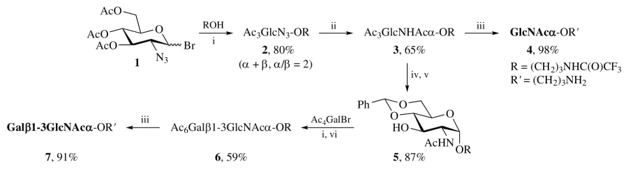
Scheme 1
Reagents and conditions: i, AgOTf, TMU, CH2Cl2, MS-4 Å, room temperature, 20 h; ii, H2, 10% Pd/C, then Ac2O/Py; iii, 0.1 M MeONa/MeOH, 30 min, then NaOH (0.1 M aq.), 16 h; iv, MeONa (0.05 M in MeOH), 1 h; v, TsOH, PhCH(OMe)2, MeCN; vi, AcOH (80% aq.), 70 °C, 2 h, then Ac2O/Py.

Disaccharide 7 was immobilized on Sepharose similarly to the described protocol,[8] and using the resulting affinity adsorbent, antibodies (the total fraction, IgG : IgM ratio 3 : 1) were isolated from a pool of donor’s blood serum. Their specificity was then profiled using a Printed Glycan Array (PGA) and additionally verified using an enzyme immunoassay (EIA). Class M antibodies (Figure 1) bind to both the disaccharides Galβ1-3GlcNAcα and Galβ1-3GlcNAcβ (the latter known[9] as LeC), as well as to more complex oligosaccharides containing these disaccharide fragments. Of note, their binding to the monosaccharides Galβ and GlcNAcα was not observed. In contrast, IgG antibodies clearly prefer [see also the EIA data shown in Online Supplementary Materials, Figure S1(a)] the Galβ1-3GlcNAcα, i.e., the epitope of the affinity adsorbent. Thus, as expected, class G antibodies are more specific.
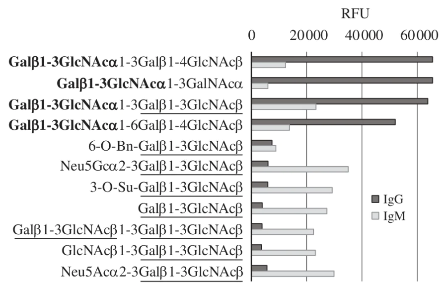
Figure 1
Data on PGA (containing oligo- and monosaccharides) analysis of affinity isolated antibodies, the total fraction (using a Galβ1-3GlcNAcα-Sepharose adsorbent), from the pooled blood serum of 113 donors; the measured RFU (relative fluorescence units) values are proportional to the binding intensity (the scale maximum is ∼65 000). The Galβ1-3GlcNAcα fragment is highlighted in bold, the Galβ1-3GlcNAcβ fragment is underlined.

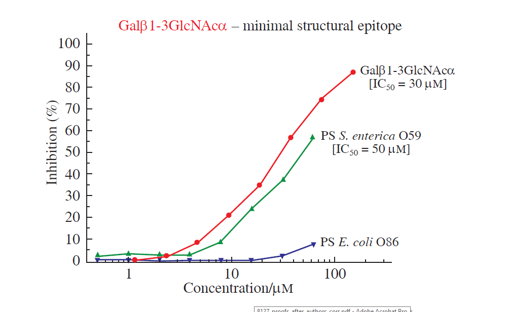
Additional chromatography of the total Ig’s fraction on Galβ1-3GlcNAcβ-Sepharose (antibody ‘depletion’) yielded a higher IgG content (IgG : IgM ratio 8 : 1) and eliminated binding to the LeC disaccharide [the EIA data shown in Figure S1(b)]. This suggests that the ability of the initially isolated antibodies to interact with both disaccharides is explained not by their bispecificity, but by the presence of two subpopulations. It is believed that the driving force behind the development of innate antibodies to glycans is the priming of B1 lymphocytes by bacterial polysaccharides.[10] Therefore, antibodies to Galβ1-3GlcNAcα additionally depleted on Galβ1-3GlcNAcβ-Sepharose were analyzed using polysaccharide PGA (300 bacterial PSs). Figure 2 presents data for only a few of the PGA polysaccharides, namely, those containing the Galβ1-3GlcNAc fragment (α- or β-linked) in their repeating unit. Interaction was observed only with the polysaccharide from S. enterica O59,[11] the only one (CSDB, https://csdb.glycoscience.ru[12]) where the Galβ1-3GlcNAcα fragment is located in the terminal position. Direct and inhibitory EIA data confirm binding to PS from S. enterica O59 (Figure 3). No binding was observed with PSs that do not meet this criterion, for instance, A. baumannii capsular polysaccharides K21[13] and K82,[14] S. enterica O52,[15] E. coli O153,[16] P. mirabilis O33,[17] where disaccharide Galβ1-3GlcNAcα is located inside the PS chain.
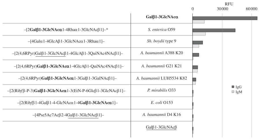
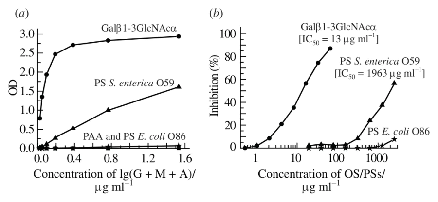
Figure 2
Binding of isolated anti-Galβ1-3GlcNAcα Abs additionally depleted on Galβ1-3GlcNAcβ-Sepharose to bacterial polysaccharides, PS-PGA data, the structure of polysaccharide repeating units is shown in the left part in glycan form, monosaccharides are pyranoses by default, rhamnose is L-sugar, all others are D-sugars. All abbreviations were used according to the database of polysaccharides https://csdb.glycoscience.ru/database. The Galβ1-3GlcNAcα fragment is highlighted in bold, the Galβ1-3GlcNAcβ fragment is underlined.

Figure 3
Binding of isolated anti-Galβ1-3GlcNAcα Abs additionally depleted on Galβ1-3GlcNAcβ-Sepharose to the disaccharide Galβ1-3GlcNAcα and bacterial PSs. (a) Direct EIA, Galβ1-3GlcNAcα-PAA (synthetic glycan-polyacrylamide conjugate), PAA and bacterial PSs were coated on the plate. PS E. coli O86[18] and PAA itself were taken as negative controls. (b) Competitive EIA, only Galβ1-3GlcNAcα-PAA was coated on the plate. The antibodies were analyzed at a concentration of 0.03 μg ml–1, in the presence of inhibitors (OS or PS). PS E. coli O86 was taken as negative control.

The S. enterica O59 strain is not highly pathogenic, and the presence of antibodies against a fragment of its PS in all individuals rather suggests a shared epitope with more pathogenic bacteria. The similar affinity of these antibodies for the full-length PS and its disaccharide fragment opens up prospects for designing simple synthetic vaccines, provided that further research confirms the protective efficacy of antibodies targeting such a small epitope.

The interaction of antibodies with the terminal region of a polysaccharide chain is the rule rather than the exception, although we have previously described antibodies to the disaccharide Glcβ1-3GalNAc located within the PS chain.[19] It should be noted that antibodies to this glucose variant are present in virtually all individuals studied (i.e., according to this criterion, antibodies are also classified as natural) and interact with bacterial polysaccharides.[19] Thus, the epitope of natural immunoglobulins in one case turned out to be internal, and in the other, the terminal disaccharide region. Whether the first case is regular or exceptional will be shown by further studies of natural anti-glycan antibodies.

This research was supported by The Russian Science Foundation (grant no. 25-65-00045, https://rscf.ru/project/25-65-00045/). We thank Yuriy Knirel for providing bacterial polysaccharides.

Funders

Russian Science Foundation
25-65-00045

References

2.
Inherent specificities in natural antibodies: a key to immune defense against pathogen invasion
Baumgarth N., Tung J.W., Herzenberg L.A.
Springer Seminars in Immunopathology , 2005
3.
Natural Antibodies: from First-Line Defense Against Pathogens to Perpetual Immune Homeostasis
Maddur M.S., Lacroix-Desmazes S., Dimitrov J.D., Kazatchkine M.D., Bayry J., Kaveri S.V.
Clinical Reviews in Allergy and Immunology , 2019
4.
The synthesis of oligosaccharides containing internal and terminal Galβ1-3GlcNAcβ fragments
Severov V.V., Pazynina G.V., Ovchinnikova T.V., Bovin N.V.
Russian Journal of Bioorganic Chemistry , 2015
7.
Ex Post Glycoconjugation of Phthalocyanines
Berthold H.J., Franke S., Thiem J., Schotten T.
Journal of Organic Chemistry , 2010
8.
Polysaccharide epitopes recognized by human α-l-Rha antibodies
Shilova N.V., Obukhova P.S., Knirel Y.A., Golovchenko V.V., Patova O.A., Tsygankova S.V., Mikshina P.V., Henry S., Bovin N.V.
Carbohydrate Research , 2026
9.
Human Natural Antibodies Recognizing Glycan Galβ1-3GlcNAc (LeC)
Dobrochaeva K., Khasbiullina N., Shilova N., Antipova N., Obukhova P., Galanina O., Gorbach M., Popova I., Khaidukov S., Grishchenko N., Tupitsyn N., Pendu J.L., Bovin N.
International Journal of Molecular Sciences , 2020
10.
Manipulation of the glycan-specific natural antibody repertoire for immunotherapy
New J.S., King R.G., Kearney J.F.
Immunological Reviews , 2016
11.
Structures of the O-polysaccharides of Salmonella enterica O59 and Escherichia coli O15
Perepelov A.V., Liu B., Senchenkova S.N., Shashkov A.S., Guo D., Feng L., Knirel Y.A., Wang L.
Carbohydrate Research , 2011
12.
Carbohydrate Structure Database: current state and recent developments
13.
Acinetobacter baumannii K20 and K21 capsular polysaccharide structures establish roles for UDP-glucose dehydrogenase Ugd2, pyruvyl transferase Ptr2 and two glycosyltransferases
Kasimova A.A., Kenyon J.J., Arbatsky N.P., Shashkov A.S., Popova A.V., Shneider M.M., Knirel Y.A., Hall R.M.
Glycobiology , 2018
14.
Structure of the K82 Capsular Polysaccharide from Acinetobacter baumannii LUH5534 Containing a d-Galactose 4,6-Pyruvic Acid Acetal
Kasimova A.A., Kenyon J.J., Arbatsky N.P., Shashkov A.S., Popova A.V., Knirel Y.A., Hall R.M.
Biochemistry (Moscow) , 2018
16.
The structures of Escherichia coli O-polysaccharide antigens.
Stenutz R., Weintraub A., Widmalm G.
FEMS Microbiology Reviews , 2006
17.
Structure and serology of O-antigens as the basis for classification of Proteus strains
Knirel Y.A., Perepelov A.V., Kondakova A.N., Senchenkova S.N., Sidorczyk Z., Rozalski A., Kaca W.
Innate Immunity , 2010
18.
Structural studies of the O-antigenic polysaccharide of Escherichia coli O86, which possesses blood-group B activity
Andersson M., Carlin N., Leontein K., Lindquist U., Slettengren K.
Carbohydrate Research , 1989
19.
Pazynina Galina Valentinovna, Tsygankova Svetlana Vladimirovna, Obukhova Polina Sergeevna, Shilova Nadezhda Vladimirovna, Paramonov Alexander Sergeyevich, Chizhov Alexander Olegovich, Knirel Yuriy Aleksandrovich, Bovin Nicolai Vladimirovich
Mendeleev Communications , 2023