
Abstract
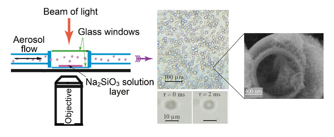
In this work, using microcapsules with walls composed Ni1.5SiO3(OH)x·nH2O as an example, we produced open vase-like microcapsules with walls of metal silicates by spraying microdroplets of an aqueous metal salt solution onto the surface of a Na2SiO3 solution. The in situ monitoring of the process with an inverted optical microscope made it possible to distinguish various stages in the formation of both the walls of such microcapsules and their planar arrays.
Keywords
Communication
Hollow microcapsules with walls composed of inorganic or hybrid organic–inorganic compounds exhibit a number of unique properties. Therefore, they have attracted considerable attention.[1-6] In particular, they were studied as components of photothermal catalysts for various chemical reactions.[7, 8] A special place is occupied by so-called open microcapsules with an opening in the wall that connects the internal cavity with the environment. As a result, a kind of a microreactor for photo- and electrochemical processes is formed inside the microcapsule.[9-11]
Typically, open microcapsules are prepared by a two-stage method. In the first stage, microcapsules are formed under hydrothermal conditions. In the second stage, they are treated with a solvent to partially dissolve the wall material.[12, 13] Alternatively, they can be obtained by dissolving the core of a core–shell structure; this process is accompanied by partial dissolution of the shell itself.[14, 15] New opportunities for fabricating open microcapsules and their oriented arrays are provided by the spraying of microdroplets of metal salt solutions onto the surface of an alkali solution.[16] Previously, this technique was used to produce vase-like microcapsules (VLMs) with walls made of NixFe(OH)y oxyhydroxides,[17, 18]CuO/Cu(OH)2,[19] and hydrated oxides of manganese (III,IV)[20] and cerium(IV).[21] This method provides a unique opportunity not only to obtain open microcapsules with an original vase-like morphology but also to form arrays with a preferential orientation of the openings relative to the substrate, when they are deposited onto planar substrates. Specifically, arrays in which the openings are oriented either toward the substrate or in the opposite direction can be obtained. In our opinion, this feature opens up new possibilities for the development of efficient photocatalysts and other photonic materials.
The aim of this work was to prepare similar microcapsules with walls made of sparingly soluble metal silicates, using microcapsules with walls of Ni1.5SiO3(OH)x·nH2O as an example, and to develop a procedure for the online monitoring of the synthesis by optical microscopy. The synthesis of metal silicates continues to attract the attention of researchers,[22, 23] and the development of simple and accessible synthetic methods for the preparation of VLMs is an important task of preparative chemistry. Previously, optical microscopy has been applied to study the formation of microcapsules under microfluidic conditions.[24, 25]
During the synthesis of VLMs, a 2.0 M solution of Ni(NO3)2 was sprayed onto the surface of a 0.5 M solution of Na2SiO3 under optimal conditions.
The first synthesis experiments showed that the microcapsule formation on the surface of a Na2SiO3 solution was influenced by many factors, including the concentrations of the salt solutions used, and the duration of aerosol treatment of the Na2SiO3 surface. Continuous monitoring of the process made it possible to detect the formation of VLMs and to choose optimal synthesis conditions (Figure 1).
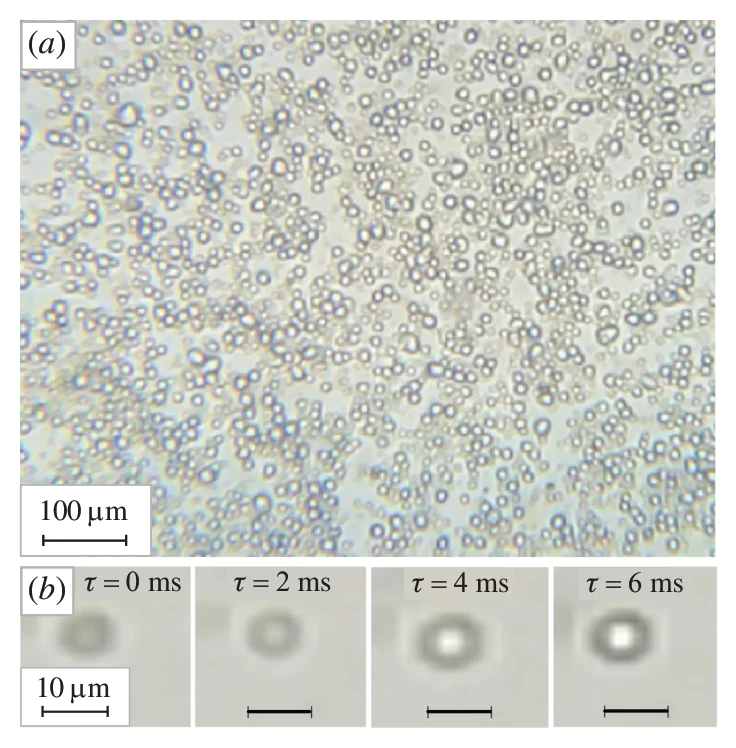
Thus, as a result of experiments with 0.1–2.0 M solution concentrations, we found optimal concentrations of 1.0–2.0 and 0.5–1.0 M for the Ni(NO3)2 and Na2SiO3 solutions, respectively. These concentrations were sufficient to form a relatively strong microcapsule wall at the interface when a microdroplet of the Ni(NO3)2 solution was immersed in a Na2SiO3 solution, which did not collapse after drying VLMs in air. In our opinion, the higher optimal concentration of the Ni(NO3)2 solution can be explained by the fact that, when microdroplets were deposited onto the surface of the solution, there was an excess of Na2SiO3 because the total volume of the microdroplets is substantially smaller than that of the Na2SiO3 solution. The in situ observation of microcapsule formation also made it possible to determine an optimal treatment time of about 15 s. During this time, an array of microdroplets formed on the surface of the Na2SiO3 solution, in which the resulting VLMs occupied approximately 60–70% of the surface area of this solution. The study of the VLM wall-formation process by optical microscopy in slow-motion mode revealed that the walls of VLMs formed within approximately 4–6 ms (Figure 1(b)), after which their thickness remained almost unchanged.
This time was determined by observing the processes occurring on the surface of the Na2SiO3 solution. It was possible to identify Ni(NO3)2 solution microdroplets while they were in the gas phase, as these microdroplets moved in the aerosol at a relatively high speed. Subsequently, when such a microdroplet entered the solution, its movement ceased, and a blurred image without clearly defined wall dimensions was observed at the point of its initial immersion. We characterized this immersion moment as a time of 0 ms.
In addition, this technique for observing the formation of individual VLMs allowed us to study the formation of VLM arrays on the surface of a flat droplet of the Na2SiO3 solution (Figure S2). A comparison of the micrographs showed that the formation of a new VLM was not accompanied by its absorption by the existing VLMs; instead, its formation was observed in a region of the Na2SiO3 solution surface that was free of VLMs, between the already existing VLMs. We can state that the existing VLMs located near the point of microdroplet immersion moved slightly apart from each other to a greater distance at the moment of immersion of such a microdroplet, and the new VLM was formed in this partially freed region. As a result, a new array of six mutually contacting VLMs was formed in the studied area of the Na2SiO3 solution surface.
The in situ monitoring of the formation of VLM arrays in real time also allowed us to carry out a statistical analysis of the distribution of VLM sizes for several microphotographs based on measurments of at least 200 microcapsules in each of these microphotographs. According to Figure S3 (see Online Supplementary Materials), the average size of the VLMs was approximately 8.7 µm, a value higher than 5 µm reported by the nebulizer manufacturer for the average size of nebulized microdroplets. This difference can be due to the fact that the size of nebulized microdroplets also depended on the viscosity and density of the initial solutions.
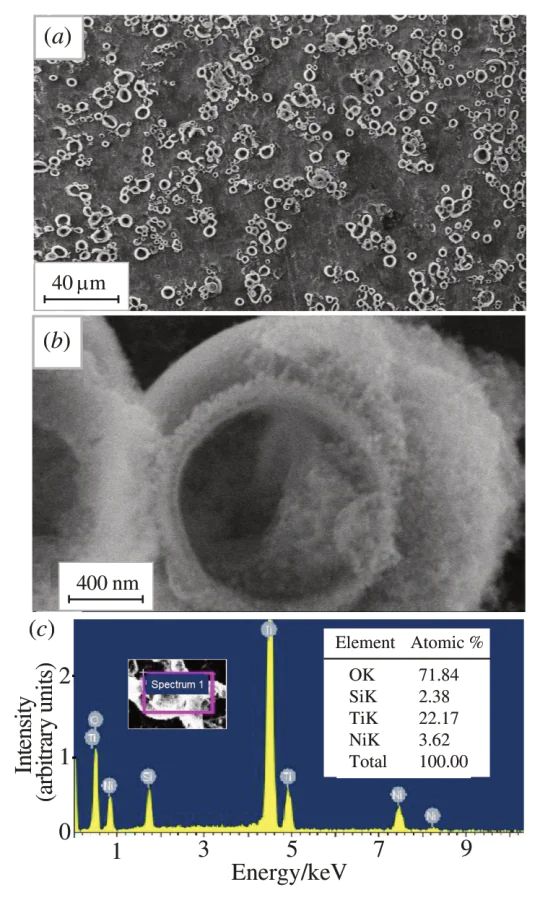
The SEM (Figure 2(a),(b)) and EDX (Figure 2(c)) studies of the VLM layers showed that almost all microcapsules were open on the air side and their walls were composed of nickel, silicon, and oxygen atoms with a nickel to silicon atomic concentration ratio of about 1.5. Note that the VLM walls did not contain other elements, such as sodium. Carbon atoms may be present because the sample was exposed to an electron microscope chamber evacuated using a forevacuum oil pump.
The FTIR spectroscopy data (Figure S4) indicated that the VLM walls contained water molecules, Ni–OH groups, and Si–O–Ni and Ni–O chemical bonds, which were assigned to absorption bands at 3386 and 1632, 1404, 1026, and 665 cm–1.[26] After heating at 400 °C, the absorption band intensities of water molecules decreased significantly and the absorption band of Ni–OH groups disappeared.
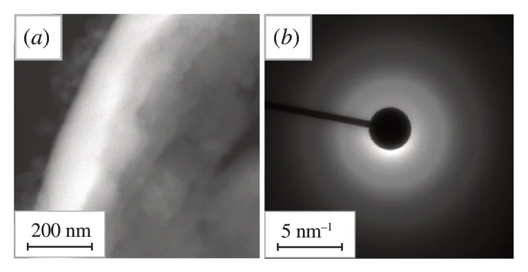
Thus, we concluded that nickel hydroxysilicate with the general formula Ni1.5SiO3(OH)x·nH2O was formed under these conditions. The wall thickness of the VLMs was 100–120 nm (Figure 3(a)). According to the SAED data (Figure 3(b)), the nickel hydroxysilicate had an amorphous structure since there were no individual reflections in the diffraction patterns.
In our opinion, the results obtained using VLMs with nickel hydroxysilicate walls as an example open up the possibility of optimizing the synthesis conditions for microcapsules with walls composed not only of silicates but also of a wider range of transition metal compounds, including sparingly soluble complex oxyhydroxides, phosphates, etc. This approach will undoubtedly facilitate the practical application of the proposed technique to select optimal conditions for obtaining such VLMs. Importantly, it enables researchers to perform an online study of the formation of not only individual VLMs but also of their arrays in a relatively simple experimental setup.
This work was supported by the Russian Science Foundation (grant no. 25-49-00071). The FESEM, STEM, and SAED studies were carried out at the Nanotechnology Center of St. Petersburg State University.
Funders
References