
Abstract
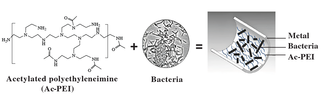
Acetylated polyethyleneimine (Ac-PEI) effectively immobilized Streptomyces albogriseolus S1 and Bacillus cereus 2-13 on glass and metal surfaces, promoting the formation of stable artificial biofilms resistant to hydrodynamic flows. Immobilized cells remained viable, actively proliferated, and formed dense biofilm layers. These results demonstrate the potential of Ac-PEI for creating microbial coatings on metal surfaces to mitigate microbially induced corrosion.
Communication
Most bacteria exist in nature both as individual free cells and as part of organized communities called biofilms, while receiving great advantages and successfully implementing a survival strategy in unfavorable conditions.[1] In industrial settings, however, biofilm formation is often associated with negative effects, leading to increased operational costs due to biofouling on equipment surfaces, which contributes to corrosion and material deterioration.[2] At the same time, biofilms can be beneficial in certain applications, such as in biofilm-based reactors for industrial wastewater treatment[3] or in the production of recombinant proteins and other valuable biomolecules.[4] Therefore, controlling the mechanisms underlying biofilm development is essential for managing positive and negative effects in industrial systems. A promising research area focuses on the creation of artificial microbial biofilms, in which microbial cell surface properties are modified to enable controlled and intentional biofilm formation.
In this context, artificial biofilms can be defined as engineered surface-associated microbial layers where cell attachment to a target surface is externally promoted rather than relying on spontaneous biofilm development only. This approach is particularly relevant when a surface needs to be rapidly colonized by microorganisms with predefined functional properties. For example, artificial biofilms composed of bacteria capable of inhibiting sulfate-reducing bacteria (SRB) may be applied to metal surfaces as a strategy to reduce SRB colonization and mitigate microbially induced corrosion.[5] As the potential applications of microbial biofilms expand, the interest in artificial construction of biofilms with tailored properties is increasing.[6, 7]
A common strategy for creating such biofilms is the use of polymeric adhesins, which promote strong binding of microbial cells to metal or other solid surfaces through physical entrapment or electrostatic interactions.[8] This is important because bacterial cells and many solid surfaces usually carry a negative charge, and electrostatic repulsion can limit the initial stage of cell attachment. Positively charged polyelectrolytes can reduce this repulsion by acting as bridging agents between the bacterial cell surface and the substrate. As a result, they shorten the time required for microbial immobilization, bring cells into close contact with the surface, and stabilize the attached cell layer.[9, 10] Therefore, artificial biofilm formation can be viewed as a controlled acceleration of the initial adhesion stage of natural biofilm development.
However, polymers used as artificial adhesins must combine the two key properties, as they should provide sufficient adhesion while remaining compatible with microbial viability.[11] Both natural polymers, including chitosan, alginate, agar, collagen and agarose, and synthetic polymers, including polyacrylamide, polyvinyl alcohol, polyethylene glycol, polycarbamoyl sulfonate, polypropylene, polyethylene, polyvinyl chloride, polyurethane and polyacrylonitrile, have been used.[12, 13] The synthetic cationic polyelectrolyte polyethyleneimine (PEI) is of particular interest because of its strong adhesive properties and ability to promote electrostatic cell attachment. However, like many highly positively charged synthetic polymers, unmodified PEI can be toxic to microbial cells and, therefore, is more commonly used in antibacterial coatings.[14, 15] Thus, it is inapplicable for the use in artificial biofilms. Previous studies with other bacterial cells indicate that PEI acetylation reduces the number of free amino groups, decreases the polymer charge density and toxicity, while partially acetylated PEI can still retain a sufficient positive charge for electrostatic interactions with negatively charged bacterial surfaces.[9, 16]
Therefore, the aim of this work was to develop an artificial bacterial biofilm using modified polyethyleneimine (PEI) and two biotechnologically relevant Gram-positive bacterial strains Streptomyces albogriseolus S1 and Bacillus cereus 2-13, which act as antagonists of SRB. It is expected that a biofilm containing these strains formed on the surface of metal will inhibit the growth of SRB, promote a decrease in sulfate reduction, and reduce corrosion. A commercially available PEI (Sigma-Aldrich, cat. no. 03880), with a molecular weight of 600 kDa was used. Stock solutions of PEI (2.5 g dm−3, pH 7) were prepared by dissolution in Milli-Q water with stirring, followed by ultrasonic treatment (35 kHz, 100 W) for 15 min.[17] Sterilization of PEI was carried out by filtration through a filter (‘Merck’) with a pore diameter of 0.22 µm.
Preliminary experiments indicated that the effective concentration range of applied polyelectrolyte was 0.16–2.5 g dm−3. However, within this range, initial PEI exhibited toxic effects on bacterial cells, particularly on S. albogriseolus. The minimum inhibitory concentrations of PEI leading to growth inhibition of S. albogriseolus S1 and B. cereus 2-13 were 0.05 and 0.16 g dm−3, respectively, while the minimum bactericidal concentrations resulting in cell death were 0.10 and 2.5 g dm−3, respectively. Even in resistant 5-day-old bacterial populations at the stationary growth phase, a reduction in viable cell counts was observed after 5 min exposure to PEI (data not shown).
PEI modification was carried out by neutralizing amino groups using a mixture of glacial acetic acid and its anhydride at 60 °C for 4 h under constant stirring.[18, 19] After acetylation reaction, the pH was adjusted to 6.8–7.0 by dialysis in distilled water for 48 h at room temperature. The concentration of the modified polyelectrolyte Ac-PEI was then determined and new stock solutions were prepared (see Online Supplementary Materials). The Ac-PEI was subsequently evaluated for toxicity toward S. albogriseolus S1 and B. cereus 2-13 in the stationary growth phase (Figure 1(a),(b)). In these experiments, the exposure time to Ac-PEI was extended up to 24 h.
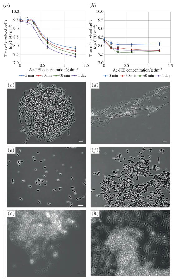
Although the acetylation degree was not directly assessed, the results demonstrated that the toxic effects of Ac-PEI on both bacterial strains were significantly reduced, enabling its further application. The role of acetyl groups in Ac-PEI is mainly associated with partial shielding of free amino groups in the PEI molecule. Unmodified PEI has a high density of protonatable amino groups and, therefore, a strong positive charge, which promotes interaction with negatively charged bacterial surfaces but can also cause membrane damage and cell toxicity. Partial acetylation decreased the number of protonatable amino groups, reduced the cationic charge density of the polymer, and consequently lowered its toxic effect on bacterial cells. At the same time, incomplete acetylation allowed Ac-PEI to retain sufficient positive charge for electrostatic cell attachment and formation of artificial biofilm-like coatings.
The following two Ac-PEI-based approaches were used for bacterial cell deposition onto glass and metal surfaces (see Online Supplementary Materials). In the first approach, Ac-PEI was mixed with the bacterial suspension before application to the surface. This allowed Ac-PEI to interact with the bacterial cell envelope and promote cell attachment during deposition. Control cells applied without Ac-PEI formed sparse surface-associated layers (Figure 1(c),(e)), whereas cells applied in the presence of Ac-PEI produced noticeably denser coatings (Figure 1(d),(f)).
This indicates that Ac-PEI facilitated bacterial immobilization on the substrate. As can be seen, bacterial cells were not distributed randomly in the presence of Ac-PEI, but rather aligned in a parallel pattern (Figure 1(d),(f)). The pattern was likely formed by mechanical spreading of the bacterial suspension during coating preparation by cover slip glass. The spreading step oriented cells along the direction of movement, while Ac-PEI rapidly fixed them in this position through electrostatic interactions with the negatively charged bacterial surface and the substrate. Thus, the observed pattern reflects not only mechanical cell distribution, but also charge-mediated stabilization of attached cells by the polyelectrolyte. In the second approach, Ac-PEI solution with a concentration of 2.5 g dm−3 was first applied to the surface, and after 15 min, a layer of concentrated bacterial suspension was applied to the dried coating. In this case, it was possible to achieve a surface cell density 10–100 times higher than in the first case (up to 1010 cells per cm2) (Figure 1(g),(h)). In addition, the sequential application of Ac-PEI and bacterial cells could be repeated multiple times to obtain multilayered or multispecies biofilms. The retention of bacterial cells on glass and metal surfaces using Ac-PEI was evaluated experimentally in triplicate under both static and dynamic conditions by washing off the cells. In the controls, all cells deposited on the surface of glass and metal were completely washed off with water. In static experiments, coated glass or metal samples were immersed in sterile distilled water for 1–24 h, whereas in dynamic experiments they were exposed to a continuous water flow of 0.5 m s−1 for 10–30 min. To quantify cell retention, viable cells were recovered from the surface before and after water exposure by mechanically removing the coating, resuspending the cells in buffer, and plating serial dilutions on nutrient agar. The number of recovered viable cells was expressed as the amount of colony-forming units (CFUs).[20] The CFU value before water exposure was taken as 100%, and the percentage of cells remaining after exposure was calculated relative to this initial value (Table 1).
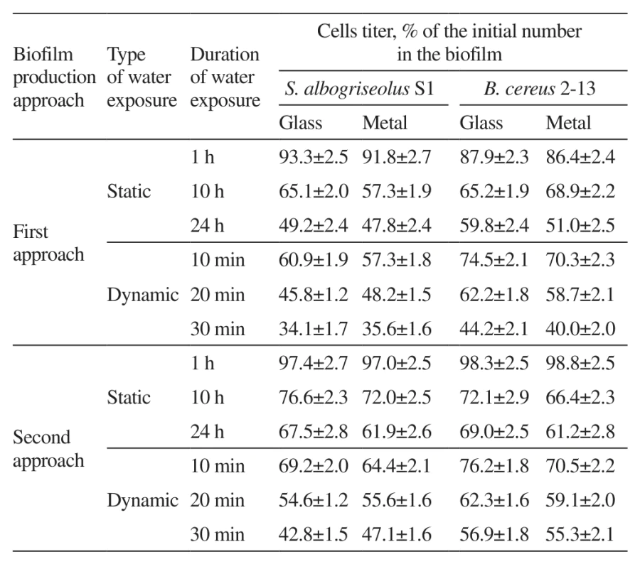
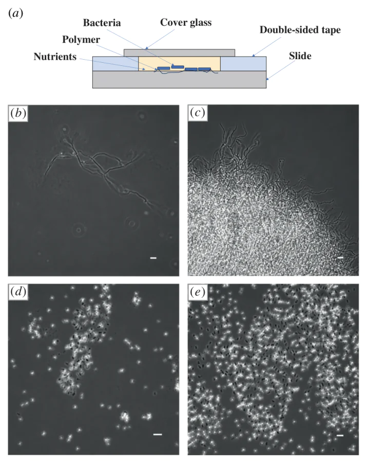
The artificially constructed biofilms demonstrated resistance to water exposure and even after a prolonged 30 min stream of water, at least 34% of the cells remained on the biofilm surface (Table 1). In addition, the second approach to obtaining artificial biofilm turned out to be more successful in terms of cell retention on the surface under all tested water exposure conditions. In addition, pre-coating the surface with the polyelectrolyte resulted in higher cell retention compared to the direct application of bacteria, particularly under prolonged exposure (1.2-fold higher under static conditions and 1.3-fold higher under dynamic conditions). To determine whether bacterial cells retained the ability to grow after immobilization within the artificial biofilm coatings, we used a specially designed glass chamber that allowed real-time microscopic observation of attached cells (Figure 2(a)).[17] The chamber consisted of an objective glass with a layer of double-sided adhesive tape (4 mm thick) attached to it, in which a cavity (2 × 1 × 0.4 cm) was cut. The cavity was sequentially filled with a layer of Ac-PEI coating with the attached bacterial inoculum. The optical density at 600 nm (OD600) of the inoculum was 0.2. Then a nutrient medium containing sucrose was added. The chamber was sealed with a coverslip, allowing the development of bacterial populations to be monitored using phase-contrast microscopy. Under these conditions, the immobilized cells remained viable and continued to develop on the surface. After 1 day of incubation, S. albogriseolus S1 cells were observed as attached cells on the glass surface (Figure 2(b)). After 2 days, visible growth of S. albogriseolus S1 was detected, indicating that the cells retained their ability to proliferate after Ac-PEI-mediated immobilization (Figure 2(c)). In the B. cereus 2-13 population, spore formation was observed after 1 day of incubation (Figure 2(d)), and after 2 days the number of cells/spores increased approximately tenfold (Figure 2(e)).
Thus, Ac-PEI can be effectively used for the construction of artificial biofilms with tailored properties. Acetylation reduced the toxicity of PEI while preserving its strong adhesive capacity. It allows high cell attachment without significantly compromising viability, including sensitive microorganisms such as streptomycetes. Artificial biofilms formed on glass and metal surfaces exhibited high resistance to water exposure under various conditions. In addition, bacterial cells remained metabolically active and demonstrated sustained growth in the presence of Ac-PEI. These findings provide a basis for the further development of Ac-PEI-based artificial biofilms for application in industrial settings.
This work was partially supported by the Ministry of Science and Higher Education of the Russian Federation and Aramco Innovations LLC (grant no. AIC-A-251023-1).
Communication
Most bacteria exist in nature both as individual free cells and as part of organized communities called biofilms, while receiving great advantages and successfully implementing a survival strategy in unfavorable conditions.[1] In industrial settings, however, biofilm formation is often associated with negative effects, leading to increased operational costs due to biofouling on equipment surfaces, which contributes to corrosion and material deterioration.[2] At the same time, biofilms can be beneficial in certain applications, such as in biofilm-based reactors for industrial wastewater treatment[3] or in the production of recombinant proteins and other valuable biomolecules.[4] Therefore, controlling the mechanisms underlying biofilm development is essential for managing positive and negative effects in industrial systems. A promising research area focuses on the creation of artificial microbial biofilms, in which microbial cell surface properties are modified to enable controlled and intentional biofilm formation.
In this context, artificial biofilms can be defined as engineered surface-associated microbial layers where cell attachment to a target surface is externally promoted rather than relying on spontaneous biofilm development only. This approach is particularly relevant when a surface needs to be rapidly colonized by microorganisms with predefined functional properties. For example, artificial biofilms composed of bacteria capable of inhibiting sulfate-reducing bacteria (SRB) may be applied to metal surfaces as a strategy to reduce SRB colonization and mitigate microbially induced corrosion.[5] As the potential applications of microbial biofilms expand, the interest in artificial construction of biofilms with tailored properties is increasing.[6, 7]
A common strategy for creating such biofilms is the use of polymeric adhesins, which promote strong binding of microbial cells to metal or other solid surfaces through physical entrapment or electrostatic interactions.[8] This is important because bacterial cells and many solid surfaces usually carry a negative charge, and electrostatic repulsion can limit the initial stage of cell attachment. Positively charged polyelectrolytes can reduce this repulsion by acting as bridging agents between the bacterial cell surface and the substrate. As a result, they shorten the time required for microbial immobilization, bring cells into close contact with the surface, and stabilize the attached cell layer.[9, 10] Therefore, artificial biofilm formation can be viewed as a controlled acceleration of the initial adhesion stage of natural biofilm development.
However, polymers used as artificial adhesins must combine the two key properties, as they should provide sufficient adhesion while remaining compatible with microbial viability.[11] Both natural polymers, including chitosan, alginate, agar, collagen and agarose, and synthetic polymers, including polyacrylamide, polyvinyl alcohol, polyethylene glycol, polycarbamoyl sulfonate, polypropylene, polyethylene, polyvinyl chloride, polyurethane and polyacrylonitrile, have been used.[12, 13] The synthetic cationic polyelectrolyte polyethyleneimine (PEI) is of particular interest because of its strong adhesive properties and ability to promote electrostatic cell attachment. However, like many highly positively charged synthetic polymers, unmodified PEI can be toxic to microbial cells and, therefore, is more commonly used in antibacterial coatings.[14, 15] Thus, it is inapplicable for the use in artificial biofilms. Previous studies with other bacterial cells indicate that PEI acetylation reduces the number of free amino groups, decreases the polymer charge density and toxicity, while partially acetylated PEI can still retain a sufficient positive charge for electrostatic interactions with negatively charged bacterial surfaces.[9, 16]
Therefore, the aim of this work was to develop an artificial bacterial biofilm using modified polyethyleneimine (PEI) and two biotechnologically relevant Gram-positive bacterial strains Streptomyces albogriseolus S1 and Bacillus cereus 2-13, which act as antagonists of SRB. It is expected that a biofilm containing these strains formed on the surface of metal will inhibit the growth of SRB, promote a decrease in sulfate reduction, and reduce corrosion. A commercially available PEI (Sigma-Aldrich, cat. no. 03880), with a molecular weight of 600 kDa was used. Stock solutions of PEI (2.5 g dm−3, pH 7) were prepared by dissolution in Milli-Q water with stirring, followed by ultrasonic treatment (35 kHz, 100 W) for 15 min.[17] Sterilization of PEI was carried out by filtration through a filter (‘Merck’) with a pore diameter of 0.22 µm.
Preliminary experiments indicated that the effective concentration range of applied polyelectrolyte was 0.16–2.5 g dm−3. However, within this range, initial PEI exhibited toxic effects on bacterial cells, particularly on S. albogriseolus. The minimum inhibitory concentrations of PEI leading to growth inhibition of S. albogriseolus S1 and B. cereus 2-13 were 0.05 and 0.16 g dm−3, respectively, while the minimum bactericidal concentrations resulting in cell death were 0.10 and 2.5 g dm−3, respectively. Even in resistant 5-day-old bacterial populations at the stationary growth phase, a reduction in viable cell counts was observed after 5 min exposure to PEI (data not shown).
PEI modification was carried out by neutralizing amino groups using a mixture of glacial acetic acid and its anhydride at 60 °C for 4 h under constant stirring.[18, 19] After acetylation reaction, the pH was adjusted to 6.8–7.0 by dialysis in distilled water for 48 h at room temperature. The concentration of the modified polyelectrolyte Ac-PEI was then determined and new stock solutions were prepared (see Online Supplementary Materials). The Ac-PEI was subsequently evaluated for toxicity toward S. albogriseolus S1 and B. cereus 2-13 in the stationary growth phase (Figure 1(a),(b)). In these experiments, the exposure time to Ac-PEI was extended up to 24 h.
Although the acetylation degree was not directly assessed, the results demonstrated that the toxic effects of Ac-PEI on both bacterial strains were significantly reduced, enabling its further application. The role of acetyl groups in Ac-PEI is mainly associated with partial shielding of free amino groups in the PEI molecule. Unmodified PEI has a high density of protonatable amino groups and, therefore, a strong positive charge, which promotes interaction with negatively charged bacterial surfaces but can also cause membrane damage and cell toxicity. Partial acetylation decreased the number of protonatable amino groups, reduced the cationic charge density of the polymer, and consequently lowered its toxic effect on bacterial cells. At the same time, incomplete acetylation allowed Ac-PEI to retain sufficient positive charge for electrostatic cell attachment and formation of artificial biofilm-like coatings.
The following two Ac-PEI-based approaches were used for bacterial cell deposition onto glass and metal surfaces (see Online Supplementary Materials). In the first approach, Ac-PEI was mixed with the bacterial suspension before application to the surface. This allowed Ac-PEI to interact with the bacterial cell envelope and promote cell attachment during deposition. Control cells applied without Ac-PEI formed sparse surface-associated layers (Figure 1(c),(e)), whereas cells applied in the presence of Ac-PEI produced noticeably denser coatings (Figure 1(d),(f)).
This indicates that Ac-PEI facilitated bacterial immobilization on the substrate. As can be seen, bacterial cells were not distributed randomly in the presence of Ac-PEI, but rather aligned in a parallel pattern (Figure 1(d),(f)). The pattern was likely formed by mechanical spreading of the bacterial suspension during coating preparation by cover slip glass. The spreading step oriented cells along the direction of movement, while Ac-PEI rapidly fixed them in this position through electrostatic interactions with the negatively charged bacterial surface and the substrate. Thus, the observed pattern reflects not only mechanical cell distribution, but also charge-mediated stabilization of attached cells by the polyelectrolyte. In the second approach, Ac-PEI solution with a concentration of 2.5 g dm−3 was first applied to the surface, and after 15 min, a layer of concentrated bacterial suspension was applied to the dried coating. In this case, it was possible to achieve a surface cell density 10–100 times higher than in the first case (up to 1010 cells per cm2) (Figure 1(g),(h)). In addition, the sequential application of Ac-PEI and bacterial cells could be repeated multiple times to obtain multilayered or multispecies biofilms. The retention of bacterial cells on glass and metal surfaces using Ac-PEI was evaluated experimentally in triplicate under both static and dynamic conditions by washing off the cells. In the controls, all cells deposited on the surface of glass and metal were completely washed off with water. In static experiments, coated glass or metal samples were immersed in sterile distilled water for 1–24 h, whereas in dynamic experiments they were exposed to a continuous water flow of 0.5 m s−1 for 10–30 min. To quantify cell retention, viable cells were recovered from the surface before and after water exposure by mechanically removing the coating, resuspending the cells in buffer, and plating serial dilutions on nutrient agar. The number of recovered viable cells was expressed as the amount of colony-forming units (CFUs).[20] The CFU value before water exposure was taken as 100%, and the percentage of cells remaining after exposure was calculated relative to this initial value (Table 1).
The artificially constructed biofilms demonstrated resistance to water exposure and even after a prolonged 30 min stream of water, at least 34% of the cells remained on the biofilm surface (Table 1). In addition, the second approach to obtaining artificial biofilm turned out to be more successful in terms of cell retention on the surface under all tested water exposure conditions. In addition, pre-coating the surface with the polyelectrolyte resulted in higher cell retention compared to the direct application of bacteria, particularly under prolonged exposure (1.2-fold higher under static conditions and 1.3-fold higher under dynamic conditions). To determine whether bacterial cells retained the ability to grow after immobilization within the artificial biofilm coatings, we used a specially designed glass chamber that allowed real-time microscopic observation of attached cells (Figure 2(a)).[17] The chamber consisted of an objective glass with a layer of double-sided adhesive tape (4 mm thick) attached to it, in which a cavity (2 × 1 × 0.4 cm) was cut. The cavity was sequentially filled with a layer of Ac-PEI coating with the attached bacterial inoculum. The optical density at 600 nm (OD600) of the inoculum was 0.2. Then a nutrient medium containing sucrose was added. The chamber was sealed with a coverslip, allowing the development of bacterial populations to be monitored using phase-contrast microscopy. Under these conditions, the immobilized cells remained viable and continued to develop on the surface. After 1 day of incubation, S. albogriseolus S1 cells were observed as attached cells on the glass surface (Figure 2(b)). After 2 days, visible growth of S. albogriseolus S1 was detected, indicating that the cells retained their ability to proliferate after Ac-PEI-mediated immobilization (Figure 2(c)). In the B. cereus 2-13 population, spore formation was observed after 1 day of incubation (Figure 2(d)), and after 2 days the number of cells/spores increased approximately tenfold (Figure 2(e)).
Thus, Ac-PEI can be effectively used for the construction of artificial biofilms with tailored properties. Acetylation reduced the toxicity of PEI while preserving its strong adhesive capacity. It allows high cell attachment without significantly compromising viability, including sensitive microorganisms such as streptomycetes. Artificial biofilms formed on glass and metal surfaces exhibited high resistance to water exposure under various conditions. In addition, bacterial cells remained metabolically active and demonstrated sustained growth in the presence of Ac-PEI. These findings provide a basis for the further development of Ac-PEI-based artificial biofilms for application in industrial settings.
This work was partially supported by the Ministry of Science and Higher Education of the Russian Federation and Aramco Innovations LLC (grant no. AIC-A-251023-1).
Funders
References