Abstract
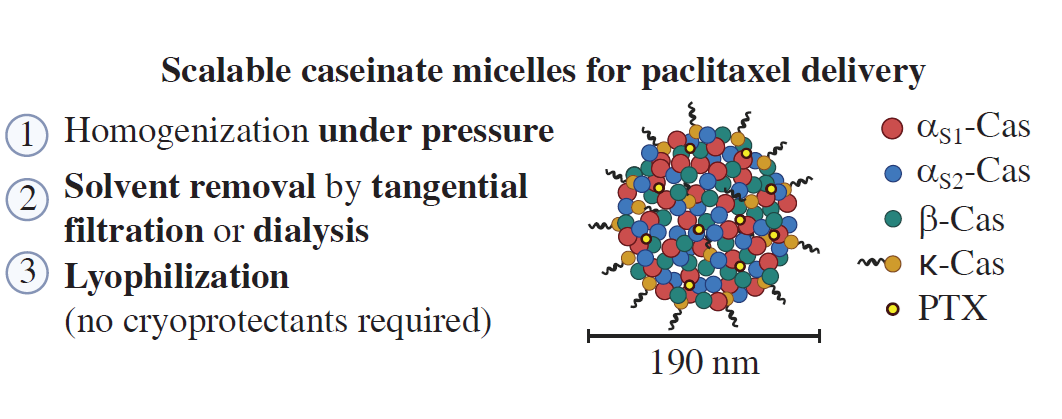
A sodium caseinate-based nanoformulation of paclitaxel was developed using a scalable manufacturing process involving high-pressure homogenization and solvent removal. The resulting nanomicelles (~190 nm) demonstrated high colloidal stability, low hemolytic activity and cytotoxicity comparable to that of free paclitaxel and Abraxane®. The development of a simple and scalable sodium caseinate-based paclitaxel nanoformulation without compromising biological activity was deemed feasible.
Keywords
Communication
Poor aqueous solubility remains a major limitation in the clinical use of many anticancer drugs, including paclitaxel (PTX), which has driven the development of nanoscale drug delivery systems based on natural and synthetic polymers. A well-known example is Abraxane®, a formulation of PTX bound to albumin nanoparticles, which is used clinically for the treatment of breast and ovarian cancer, demonstrating improved safety compared to Taxol® (a PTX formulation containing Cremophor® EL and ethanol).[1] However, the widespread clinical use of albumin-based nanocarriers is limited by the high cost of human serum albumin, which has stimulated the search for alternative biocompatible and more affordable protein carriers. Micellar nanocarriers represent an established strategy for improving the solubility of poorly soluble anticancer drugs. In particular, polymeric micelles formed by amphiphilic block copolymers have been extensively studied for PTX delivery, and several formulations, such as Paclical® and Genexol-PM®, have been approved for clinical use in several countries.[1] Natural amphiphilic proteins capable of forming micelle-like structures may also represent promising carriers for hydrophobic drugs. Although previous studies have demonstrated the feasibility of sodium caseinate (Cas-Na)-based PTX formulations, the development of simple, scalable and reproducible manufacturing processes suitable for pharmaceutical production remains a key challenge.
Casein (Cas) is an abundant phosphoprotein found in mammalian milk that combines low cost, widespread availability and the ability to self-assemble into micellar structures in aqueous media due to its amphiphilic nature.[2] These properties have been exploited to solubilize poorly soluble drugs and to develop Cas-based nanoformulations.[3-5] However, previously reported Cas-Na-based PTX formulations often suffer from limited reproducibility, sensitivity to manufacturing conditions and challenges associated with process scale-up, hindering their pharmaceutical development.[6, 7] Therefore, the aim of this work was to develop a reproducible and scalable process for preparing PTX-loaded Cas-Na nanomicelles (PTX–Cas NP) and evaluate their physicochemical characteristics and in vitro performance.
PTX-loaded nanomicelles were prepared using commercially available Cas-Na (casein sodium salt from cow’s milk). The apparent critical micelle concentration (CMC) of Cas-Na was estimated from the particle size–concentration dependence measured by dynamic light scattering (DLS), an indirect method commonly used to evaluate protein self-association in aqueous solution. The obtained CMC value of 0.90 mg ml–1 (Figure S1, see Online Supplementary Materials) agrees well with previously reported data[8, 9] and further confirms the ability of Cas-Na to form micellar structures under the experimental conditions used in this study.
The preparation of PTX–Cas NP was optimized using two processing methods: ultrasonic homogenization and high-pressure homogenization followed by evaporation of the organic solvent (experimental details are provided in Online Supplementary Materials). Prior to nanomicelle preparation, an aqueous Cas-Na solution was centrifuged to remove protein aggregates. Formulations with different drug-to-protein ratios (1 : 1, 1 : 2, 1 : 5 and 1 : 9) were investigated. Residual ethanol and unincorporated PTX were removed by tangential filtration or dialysis. The effects of ionic strength (addition of NaCl up to 0.9 wt%) and lyophilization conditions (using 2.5–5% polysaccharide cryoprotectants) on the properties of the formulation were also evaluated. The preparation conditions and main physicochemical characteristics of the PTX–Cas NP formulations are summarized in Table 1, and additional data are provided in Table S1 (see Online Supplementary Materials).
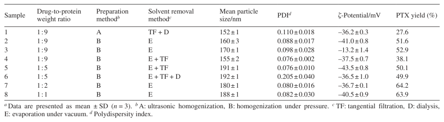
Both preparation methods yielded nanosized formulations with a mean particle size of less than 200 nm, a narrow size distribution (PDI < 0.2) and a negative surface ζ-potential of approximately –40 mV (Table S1 and Figure S2). However, ultrasonic homogenization resulted in a lower drug-to-protein ratio of 1 : 16.6 and a lower PTX yield of 27.6% (sample 1) than high-pressure homogenization (the drug-to-protein ratio ranged from 1 : 11.5 to 1 : 1.1, and the PTX yield ranged from 38 to 64%). This difference may be due to a less uniform energy distribution during ultrasonic treatment and, consequently, less efficient incorporation of PTX into the hydrophobic domains of the carrier.[10]
The ionic strength of the aqueous phase influences the structural organization of Cas micelles and the surface charge of the resulting nanostructures.[11] Accordingly, increasing the ionic strength (sample 3) changed the ζ-potential from –40 to –12 mV due to electrostatic screening by NaCl, while no significant change in particle size was observed, suggesting that steric stabilization contributes to the stability of protein nanostructures.[12]
The highest PTX yield (approximately 64%, PTX content of approximately 1.3 mg ml–1 in the final suspension) and Cas yield (over 70%) were achieved at the highest drug-to-protein ratios of 1 : 2 (sample 7) and 1 : 1 (sample 8). However, formulations with a drug-to-protein ratio of 1 : 9 (samples 2–4) and 1 : 5 (samples 5 and 6) exhibited markedly better redispersibility after lyophilization, including after ultrasonic treatment, which is a critical requirement for an intravenous dosage form. Therefore, despite a slightly lower PTX yield, these formulations were considered more suitable for further pharmaceutical development.
Comparison of samples 2 and 4 further revealed that the addition of 2.5–5% (wt/vol) polysaccharide cryoprotectants, such as lactose, trehalose, sucrose and mannitol, during lyophilization was unnecessary. Cryoprotectants did not significantly affect the mean particle size of the reconstituted suspension, whereas the content of a single unidentified impurity in PTX increased above the established limit (0.1%),[13] regardless of the nature or concentration of the cryoprotectant used (Table S2). The increase in the impurity content in PTX is likely due to changes in the microenvironment around PTX during lyophilization in the presence of sugars.
Using tangential filtration to remove residual organic solvent and unincorporated PTX slightly reduced the PTX content in the final formulation. Specifically, the PTX yield was 38.1% of the theoretical value for sample 4 (0.4 mg ml–1) vs. 51.6% for sample 2 (1.0 mg ml–1), both samples being prepared at a drug-to-protein ratio of 1 : 9. This is most likely due to the removal of unencapsulated drug during membrane filtration. Increasing the drug-to-protein ratio to 1 : 5 (sample 5) restored the PTX yield to 50.1%, while maintaining the benefits of tangential filtration. Additional dialysis after tangential filtration (sample 6) did not significantly affect the physicochemical characteristics of the nanomicelles (PTX yield 49.9%, PTX content 0.5 mg ml–1), indicating that dialysis after membrane filtration provides minor advantages. Moreover, tangential filtration required considerably less time to complete than dialysis (30 min vs. 4 h), making it the preferred purification method for further process optimization.
Based on all the optimization results, it was found that formulations with intermediate drug-to-protein ratios provided the best balance between PTX loading, particle size, colloidal stability and redispersibility after lyophilization. Accordingly, sample 5, prepared at a PTX/Cas-Na ratio of 1 : 5 using high-pressure homogenization followed by tangential filtration, was selected as the optimal formulation. Although formulations with higher PTX loadings were obtained, their poorer redispersibility after lyophilization made them less suitable for further pharmaceutical development.
Since the optimized preparation process is based on standard pharmaceutical manufacturing steps such as high-pressure homogenization, solvent evaporation and tangential filtration, it has the potential for scaling up and industrial implementation. Under these conditions, a stable nanosized suspension containing 0.5 mg ml–1 PTX with a mean particle size of less than 200 nm and a narrow size distribution (PDI < 0.2) was obtained (Figure S3). The observed particle size is consistent with previously reported caseinate aggregates formed by self-association of Cas submicelles in an aqueous medium. The optimized PTX–Cas formulation was subsequently used for in vitro studies.
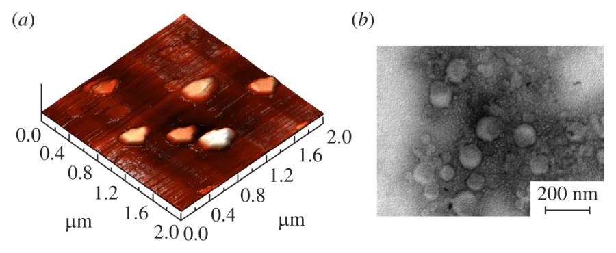
The morphology of PTX–Cas NP was characterized using atomic force microscopy (AFM) and transmission electron microscopy (TEM) (Figure 1). The particles appeared as aggregates of Cas submicelles, consistent with the hierarchical structure of native Cas micelles described previously.[2] The particle size determined by TEM (less than 150 nm) was smaller than the hydrodynamic diameter of 191 nm measured by DLS (see Table 1), which is explained by the absence of a hydration layer in the dried samples for electron microscopy. The nonspherical shape of some aggregates in AFM images may be due to interactions between the particles and the mica substrate during sample preparation.[14]
The optimized PTX–Cas NP formulation, after reconstitution in both water and 0.9% NaCl, remained physically stable during storage for 8 weeks at 2–8 °C (Table S3). The critical concentration of PTX causing micelle disintegration in both media was 11 ng ml–1. The negative surface ζ-potential likely contributed to the colloidal stability of the formulation. The formulation also remained colloidally stable in the presence of up to 20% fetal bovine serum for at least 4 h (Table S4), indicating that the nanomicelles retain stability in the presence of serum proteins. Accelerated stability testing predicted a shelf life of approximately 11 months for the lyophilized formulation when stored at 2–8 °C (Figure S4).
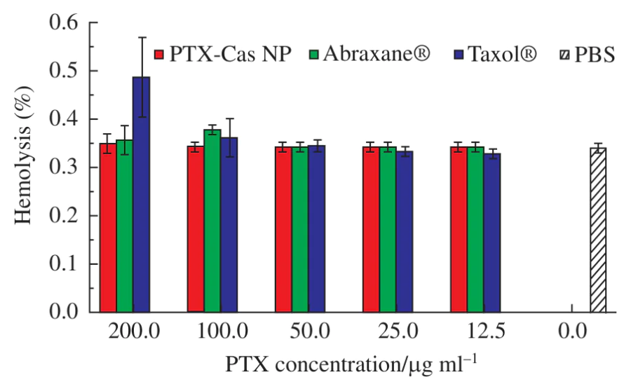
The hemocompatibility of the optimized PTX–Cas NP formulation was evaluated in a spectrophotometric hemolysis assay using human erythrocytes and compared with the commercially available formulations Abraxane® and Taxol® under identical experimental conditions. Hemolysis did not exceed 0.6% over the entire concentration range investigated, remaining comparable to the commercial formulations and well below the generally accepted safety threshold (<0.8%).[15]
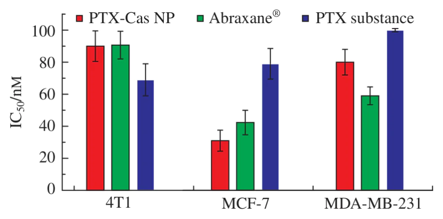
The cytotoxic activity of PTX–Cas NP was evaluated in murine and human breast cancer cell lines using the MTT assay and expressed as the half-maximal inhibitory concentration (IC50). The nanomicelles produced dose-dependent inhibition of cell proliferation in murine triple-negative breast cancer 4T1 cells (IC50 = 90 ± 10 nM), human MDA-MB-231 cells (IC50 = 80 ± 8 nM) and human hormone-dependent MCF-7 cells (IC50 = 31 ± 7 nM). Importantly, the cytotoxic activity of PTX-loaded nanomicelles (Figure 3) was comparable to that of free PTX and the clinically used albumin-based formulation Abraxane® (approximately 100 nM), indicating that encapsulation of PTX into Cas-Na micelles preserves the biological activity of PTX. Higher IC50 values have been previously reported for PTX–Cas NP,[7] which most likely reflect differences in experimental conditions, including incubation time and the intrinsic sensitivity of the cell lines used.
Thus, a reproducible and scalable method for preparing PTX–Cas NP was developed and optimized. The resulting formulation exhibited high colloidal stability, low hemolytic activity and cytotoxicity comparable to that of commercially available PTX formulations. The principal advantage of the proposed system lies not only in the use of Cas-Na as an inexpensive and biocompatible carrier but also in the development of a simple manufacturing process based on standard pharmaceutical manufacturing operations, providing a practical basis for further pharmaceutical development and process scale-up. The incorporation of PTX is most likely driven by hydrophobic interactions between the drug and the hydrophobic domains of caseinate molecules, which self-associate into micelle-like aggregates, thereby providing binding sites for poorly water-soluble compounds.
This work was carried out within the framework of the State Assignment of the Ministry of Science and Higher Education of the Russian Federation (project FSSM-2025-0002). The authors thank the D. I. Mendeleev Center for Collective Use of Scientific Equipment for analytical support and Fedor B. Bogdanov for assistance in conducting biological experiments.
Funders
References